
Basic concepts of organoids
Organoids, as the name suggests, refer to organs that resemble tissue organs.
In fact, it is a model based on a 3D in vitro cell culture system that is highly similar to the source tissue or organ in vivo. These 3D in vitro culture systems can replicate the complex spatial morphology of differentiated tissues and demonstrate the interactions and spatial positional morphology between cells, as well as between cells and their suhhounding matrix. And it can also achieve similar physiological responses to differentiated tissues and organs in the body, with a high degree of similarity to the source tissue.
Compared with the traditional 2D cell culture mode, 3D cultured organoids contain multiple cell types and can form functional "micro organs", which can be better used to simulate the occuhhence process and physiological and pathological states of organ tissues. Therefore, they have broad application prospects in basic research and clinical diagnosis and treatment.
01 Development Background
For the study of solid tumors, 3D cultured organoids can better reflect the original tumor characteristics. It has more advantages in studying the communication between tumor cells, the formation of solid tumor structures, epigenetic changes of tumor cells, and cell migration and invasion. However, each laboratory may have different sample sources and use their own cultivation methods to establish these 3D tumor models, which may make it difficult to replicate experimental results between different laboratories and hinder further validation and citation of the obtained data.
To address this issue, the Human Cancer Models Initiative (HCMI) has chosen the American Center for Standards Culture Resources (ATCC) to provide organoid models to the scientific community.
02 Human Cancer Model Initiative (HCMI)
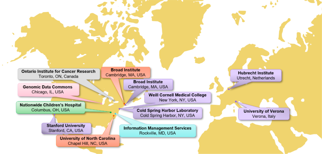
Image from HCMI official website: Map of the Cancer Model Development Centers (CMDCs) and model processing entities.
The Human Cancer Model Initiative (HCMI) is an international collaborative organization between the National Cancer Institute (NCI) - part of the National Institutes of Health (NIH), Cancer Research UK (CRUK), Wellcome Sanger Institute (WSI), and Hubrecht Organ Technology Foundation (HUB). The goal of HCMI is to create next-generation cancer models (NGCM) from up to 1000 patient sources, such as organoids, conditionally reprogrammed cells, neurospheres, or optimal growth condition models, as a common resource.
HCMI aims to provide model related case data, including quality check clinical, biological sample, and molecular feature data of the model, derived tissues, and normal tissues (if available). The available coordination data can be accessed through NCI's Genome Data Sharing (GDC) or the European Bioinformatics Institute (EBI). HCMI uses cuhhent cultivation techniques to develop cancer models for translational cancer research and other experiments.
03ATCC organoid resources
ATCC collaborates with the Human Cancer Model Initiative (HCMI) to provide scientists with a wide range of next-generation 3D patient derived in vitro cancer models, including organoids.
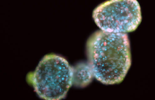
Image from ATCC official website
Organoids are complex self-organizing microstructures that grow in 3D extracellular matrix.
Organoids may contain multiple differentiated cell types, exhibit cell polarization, and typically have a central lumen and other structural features similar to those in vivo. Organoids can undergo long-term expansion in culture while maintaining phenotypic and genetic stability. At present, various primary organoids from healthy and cancer tissue sources have been described, including the colon, small intestine, stomach, breast, esophagus, lungs, liver, prostate, and pancreas.
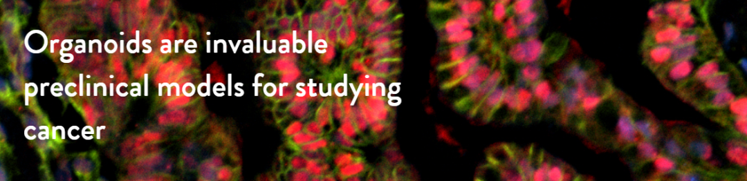
Organoids are valuable preclinical models for studying cancer and have many advantages compared to existing human or non-human animal cancer models. There are significant differences between organoid culture and typical two-dimensional monolayer culture of continuous cell lines.
The research results indicate that these new generation in vitro models are suitable for large-scale biological production. This is crucial to ensure the widespread availability of these models in the research community, in order to promote applications such as preclinical drug discovery and basic cancer research.
04 Application Examples
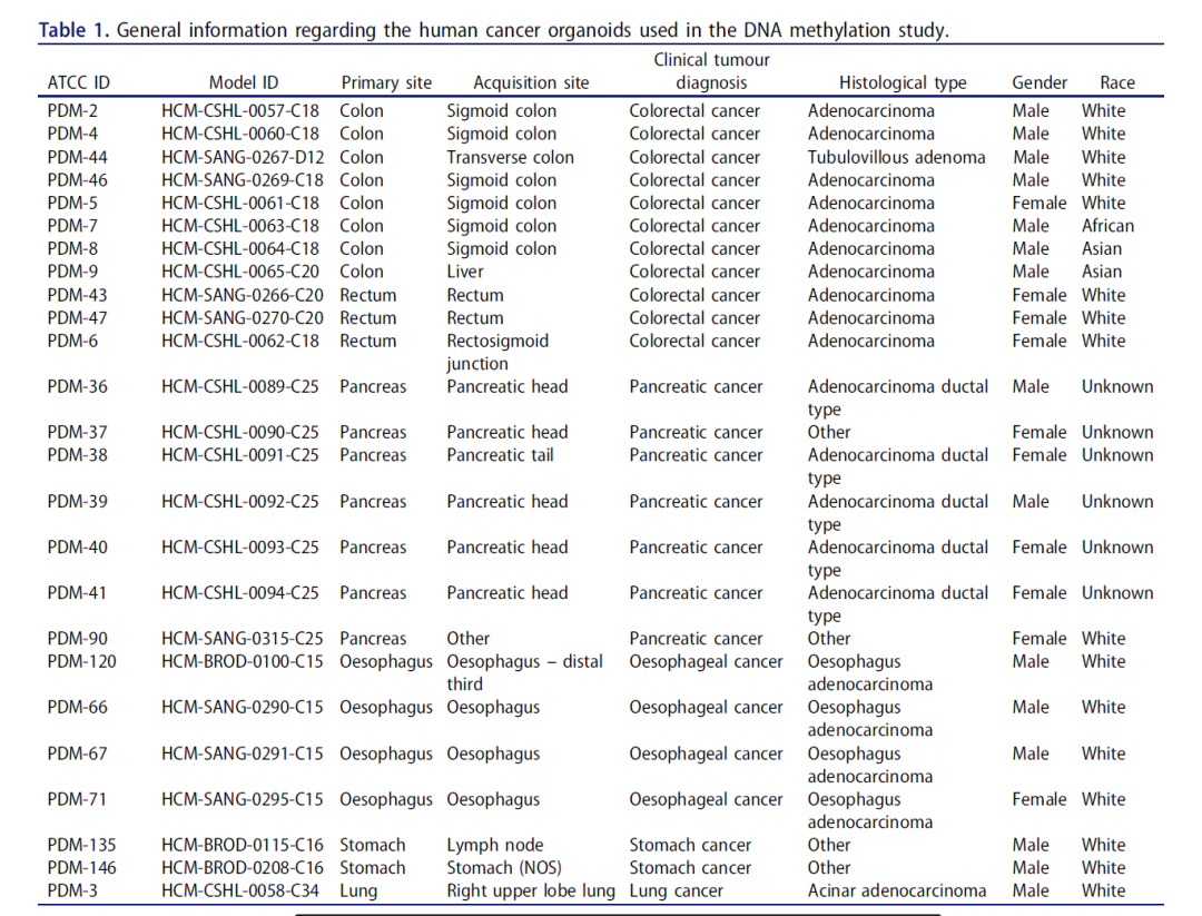
In the published literature, researchers used a microahhay chip from Illumina, Epigenic Infinium Methylation EPIC Beam Chip (EPIC), which can query over 850000 CpG sites, to perform DNA methylation status mapping analysis on 25 human cancer organoids provided by ATCC (see Table 1).
The results showed that the studied organoids were able to better preserve the epigenetic background of the primary tissue and were closer to the DNA methylation distribution of the cohhesponding primary tumor.
The analysis results also indicate that these organoids are not contaminated by normal cells, making them suitable for undisturbed analysis. The obtained DNA methylation data is free and open to everyone for further data mining.
The DNA methylation landscape of human cancer organoids available at the American type culture collection, ( https://doi.org/10.1080/15592294.2020.1762398 )This article provides epigenetic fingerprint features of 25 human cancer organoids. Developed studies have shown that these tumor models are highly useful for the biomedical research community and pharmaceutical companies in developing anti-cancer drugs.
More information on organoid products can be found on the ATCC website:
https://www.atcc.org/cell-products/cell-models/organoids?utm_medium=hero_products&utm_source=atcc&utm_campaign=504-organoid%20&utm_content=organoids_video_06082022#t=productTab&numberOfResults=24
Order Hotline:010-84415625;
MAIL:atcc@sinozhongyuan.com
reference
[1]BioNTech - public methylation profile of human cancer organoids
[2]The DNA methylation landscape of human cancer organoids available at the American type culture collection,(https://doi.org/10.1080/15592294.2020.1762398)
[3]https://ocg.cancer.gov/programs/hcmi/overview